Support breakthrough brain imaging research for neurological conditions
Using advanced measurements in scans for better brain imaging.
Magnetic resonance imaging (MRI) is a continually evolving technology, probing information from atoms and molecules to measure brain physiology.
Improving brain imaging is key to getting a clearer picture of the brain. Imaging is the only way we can see inside the brain without surgery and so is critical for monitoring disease.
Why funding is needed
Paying for MRI scans for imaging research
A 15-minute MRI scan costs £100. Typically, 100 scans would be required to provide the proof of concept needed to start to collect this information in clinical practice for patient benefit.
Funding an imaging PhD student
For postgraduate researchers, it costs £100,000 to carry out PhD research into this area. Examples of projects in this field include developing new MRI techniques to help us better understand neuroinflammation in the human brain, and studying changes to the blood supply in vascular dementia to see how they affect the brain.
More information about brain imaging research
Imaging advances often work alongside other technologies, like brain stimulation and focused ultrasound, which help us target new treatments more accurately.
With extra funding, we can buy new equipment, come up with new ways to measure things using our current tools, and train people with the skills needed to turn these measurements into real benefits for patients.
We can also attract physics and engineering graduates into the field to invent robust new measurements in line with our evolving understanding of disease mechanisms.
These measurements will allow us to diagnose, predict and monitor disease processes and provide critical information when testing the effectiveness of new treatments.
A 15-minute MRI scan can help us acquire the most advanced measurements in patients. This will allow us to test whether the new measurements we’re taking can help us target treatments more effectively and track disease progress.
New measurements in brain imaging
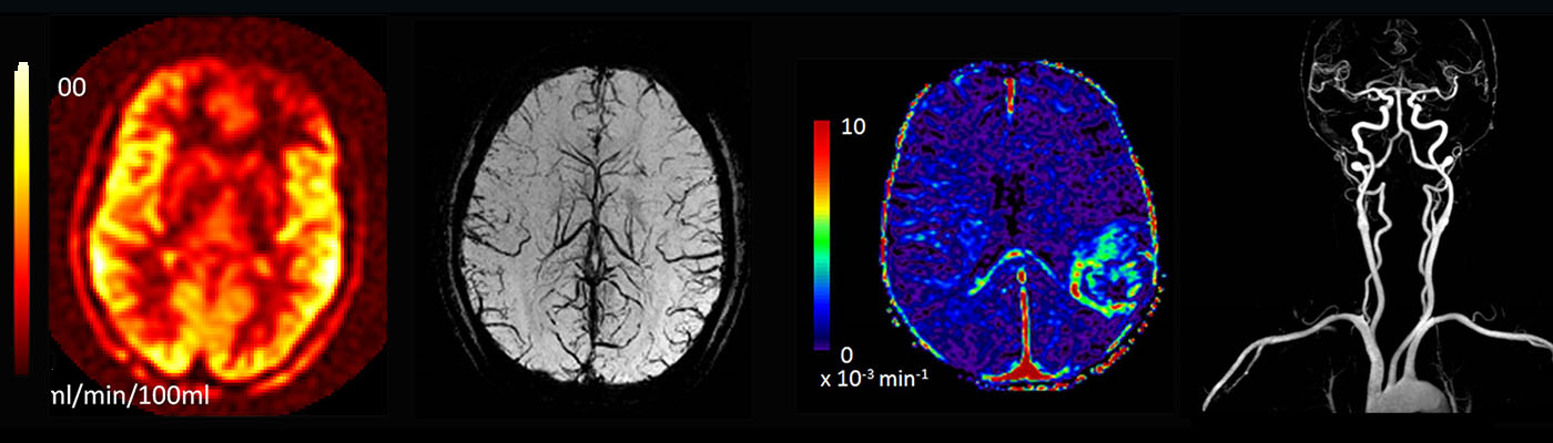
Arterial spin labelling, amide proton transfer and quantitative susceptibility mapping are examples of new measurements that we have developed over the last 10 years, which are now starting to be used in clinical practice.
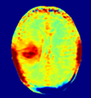
Amide Proton Transfer: This looks at the increased cell division in active tumour cells. It could be really useful for monitoring patients with brain tumours, making sure their treatment is as effective as possible.
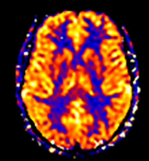
Arterial Spin Labelling: This measures blood flow in the brain and could help predict how cognitive decline might progress in people at the early stages of dementia. It could also help identify which patients are most likely to benefit from new dementia treatments.
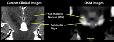
Quantitative Susceptibility Mapping (QSM): This technique is sensitive to iron buildup in the brain, and can be very helpful in identifying the sub-thalamic nucleus – a part of the brain that is targeted in deep brain stimulation for Parkinson’s disease.
Contact us
If you have any questions, please email us at GJBRC@manchester.ac.uk.
Address
Geoffrey Jefferson Brain Research Centre
Clinical Sciences Building
Northern Care Alliance
NHS Foundation Trust
Salford
M6 8FJ
Email: GJBRC@manchester.ac.uk
Connect with us