MRC award to unravel the role of the thalamus in memory
Talking about the project Alex said “I am thrilled and humbled to receive funding from the MRC (£800k +)…I am so grateful for this opportunity and eager to start working on this project!”
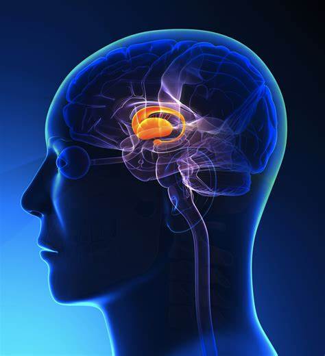
The thalamus, a brain structure deep inside the brain, includes multiple nuclei called anterior, mediodorsal, ventral (further separated into anterior, lateral and posterior) and pulvinar thalamic nuclei. The specific contribution of these structures in memory retrieval and the way they communicate with other critical structures of the memory network remain understudied especially in humans. Damage to some of these structures is linked to memory disorders, which means they should support functions related to memory. However, whether this damage disrupts processes directly related to memory retrieval or can be explained by other cognitive dysfunction is disputed. It is also disputed whether damage in these nuclei prevents patients recollecting their memory or, in addition, it affects their feeling of familiarity for previously experienced things. The
main reasons these questions are still outstanding in the memory literature, relate to key methodological considerations, including small number of participants – especially patients with damage in the thalamus – and insufficient measures of cognition and memory in previous studies.
In the proposed project, the team will select 40 patients with memory problems as a result of thalamic stroke resulting in reduced volumes in these structures. State-of-the-art brain imaging will be used to measure these structural volumes, as well as the size of fibre tracts and spontaneous connectivity mediated by the thalamic nuclei. Tests of cognitive function and memory, including tests of familiarity and recollection for different types of stimuli, such as scenes, objects and words, will be given to patients and healthy participants. Functional brain imaging in healthy participants, while undergoing task-based fMRI (2 experiments) will also be used to explore whether these structures contribute to memory retrieval in communication with other structures of the memory network. These functional brain measures will be also complemented by synchronised recording of eye movements to explore the way the various thalamic structures (and especially the ventrolateral and pulvinar areas) integrate visual information in the service of memory. By relating memory and cognitive tests with brain imaging data, we will determine how the various thalamic structures function in the different types of memory, whether the role of the thalamus in memory can be explained by other higher cognitive functions and whether specific thalamic nuclei are responsible for integrating and communicating memory signals. The findings will fundamentally change the way the role of the thalamus is described within the brain’s memory networks.


Address
Geoffrey Jefferson Brain Research Centre
Clinical Sciences Building
Northern Care Alliance
NHS Foundation Trust
Salford
M6 8FJ
Email: GJBRC@manchester.ac.uk
Connect with us
0 Comments